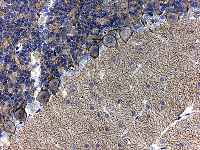
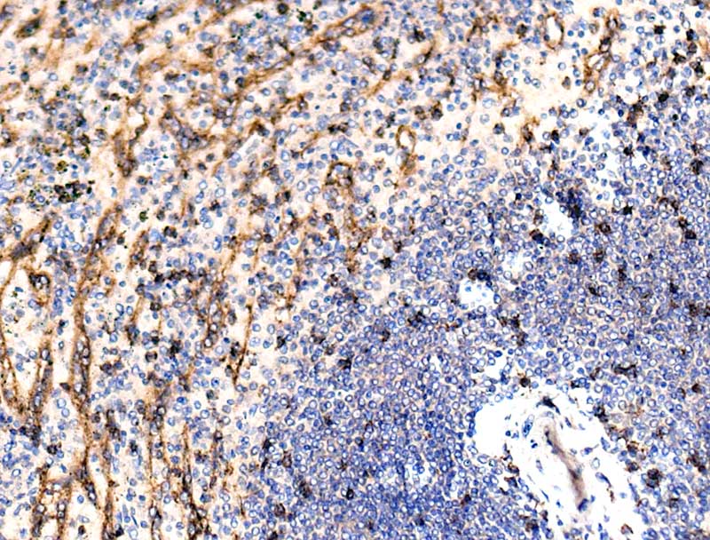
Identical positivity to that seen in rat cerebellum.
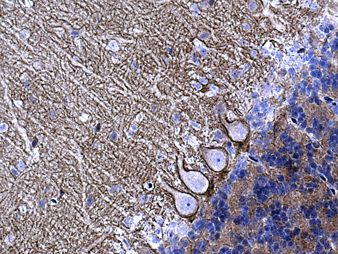
Interestingly, the most intense positivity is seen on the membrane of the soma of Purkinje cells; this extends a short distance along the primary dendrite and then appears to cease to be expressed.
Positivity on the membranes of the Purkinje cell secondary dendrites and also intense positivity apparently on the membranes of the Purkinje cell bodies
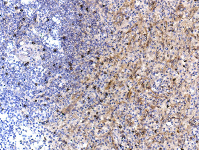
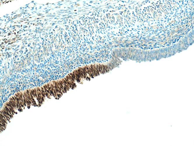
Pattern of positivity is identical to that shown by HPA. Intensely stained individual cells are scattered throughout the white/red pulp. The cells lining the sinusoids also appear positive: according to Dako spec sheet, this is specific