Monthly Archives: May 2015
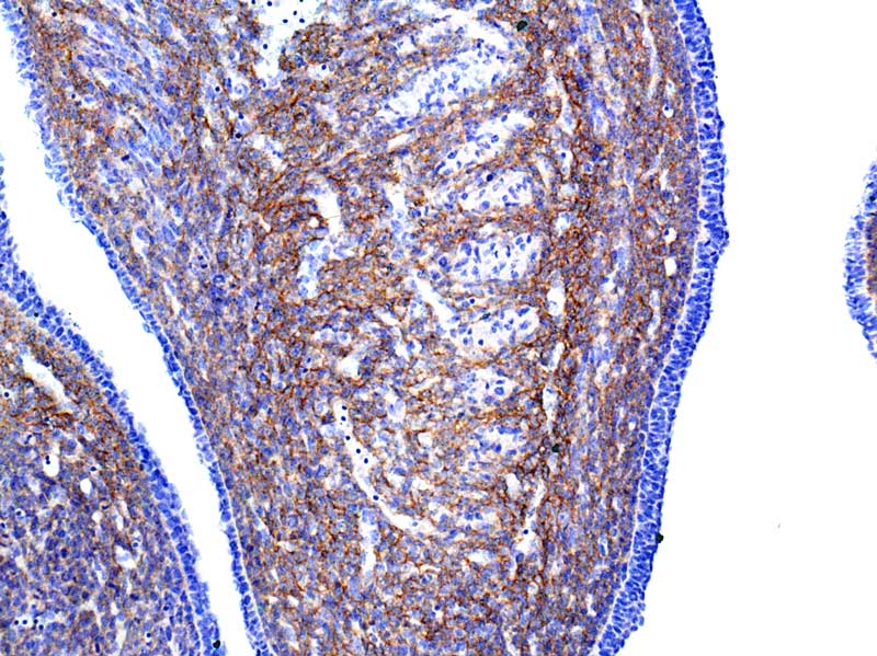
pdgf receptor alpha in mouse e18 : FFPWS using R&D systems AF1062
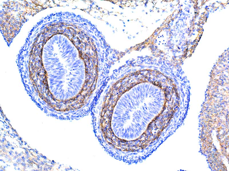
pdgf receptor alpha in mouse e18 intestine : FFPWS using R&D systems AF1062
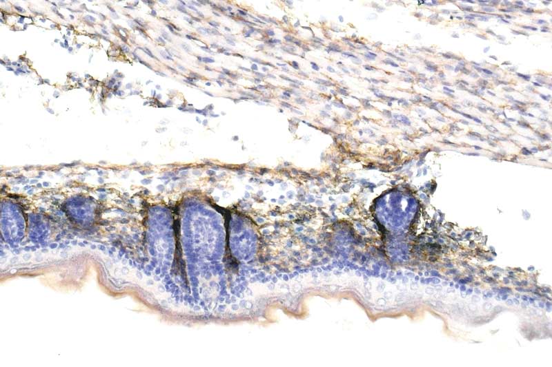
pdgf receptor alpha in mouse e18 skin : FFPWS using R&D systems AF1062
Cleaved caspase 3 in mouse neurons: FFPWS using Cell Signalling’s 9661
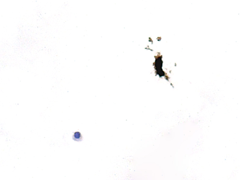
As Matrigel IF post but, using DAB. Note viable, non-apoptotic cell on the left.
Cleaved caspase 3 in mouse neurons: FFPWS using Cell Signalling’s 9661
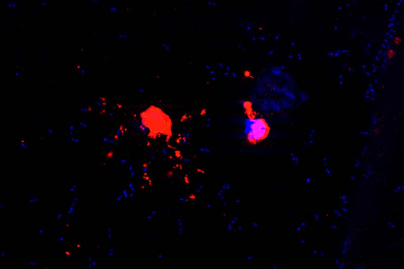
Primary cultured neurons induced to apoptose by addition of Amyloid beta. The cell on the left is fragmenting and contains no nucleus ( negative for Hoechst). The cell on the right is in early stages of apoptosis as it still has a Hoechst-stained nucleus.
Cells were grown in matrigel. Fixed and processed to Pwax.
Note many Hoechst-positive small objects: most likely the culture was contaminated and these are bacteria.
Comments welcome.
Connexin 43 and HCN4 in mouse heart: FFPWS using Ab11370 and ab69054, respectively.
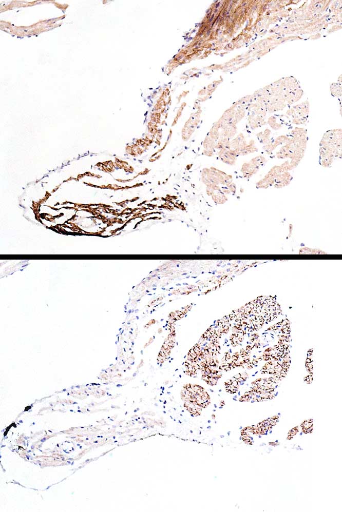
Upper image ( anti HCN4) shows strong SA node positivity. Sure, a moderate b’ground positivity that could be lowered on further dilution.
Lower image ( anti Connexin 43 ) shows negative staining of SA node but strong positivity of intercalated discs of cardiomyocytes.
CACNA2D1 (calcium channels) in rat spinal cord: FFPWS comparing HPA008213 ( atlas) and ab2864 ( abcam)
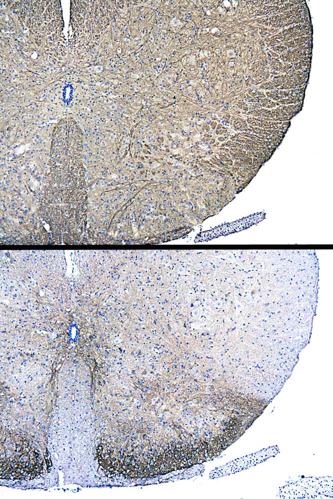
I was informed that CACNA2D1 ( upper image) and ab2864 ( lower image) targeted two different proteins but, Abcam ‘s alternative names list, for ab2864, includes CACNA2D1.
So, if these two antibodies target the same protein, why do they give very different results? Other images of CACNA2D1/HPA008213 I have uploaded show “excellent” positivity in myelinated nerve fibres.
However, I understand that CACNA2D1 should be present in the superficial laminae of the spinal cord. Abcam’s ab shows this, Atlas’ ab does not.
Comments/elucidation most welcome.
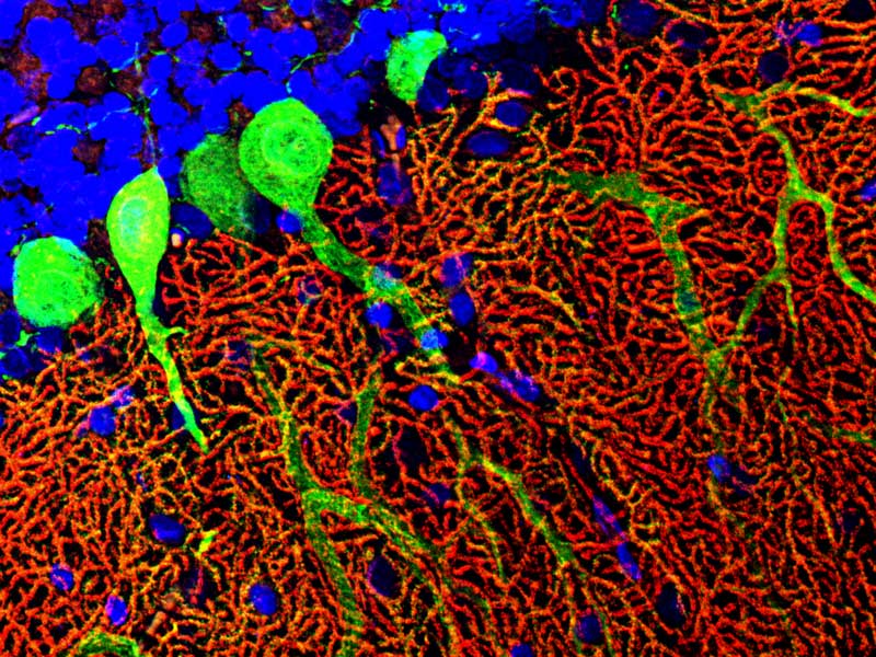
Dag lipase alpha (red – inhouse ab) and calbindin D ( green – Sigma C9848)