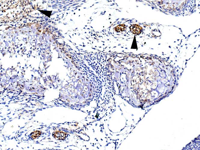
Abcam state this Ab as being non-IHC-P reactive.
The only positivity I see ( in IHC-P) is in nerve fibres of developing mouse ( arrowheads indicate positivity).
Spurious?
Abcam state this Ab as being non-IHC-P reactive.
The only positivity I see ( in IHC-P) is in nerve fibres of developing mouse ( arrowheads indicate positivity).
Spurious?
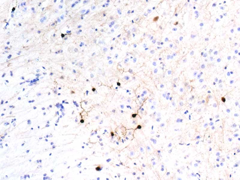
Positive cells in hypothalamic region.
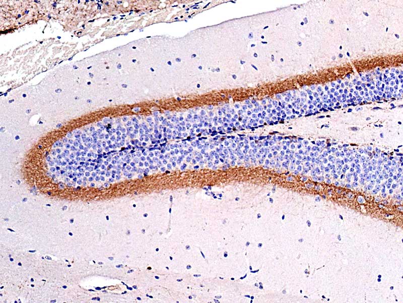
Supra-pyramidal blade ( is this correct?) shows strong positivity
A subset of neurones show a strong dense cytoplasmic and a punctate process positivity.
Why only a subset of neurones?
Amplified using BA-6000 then stABC-px and visualised by in-house DAB formulation.
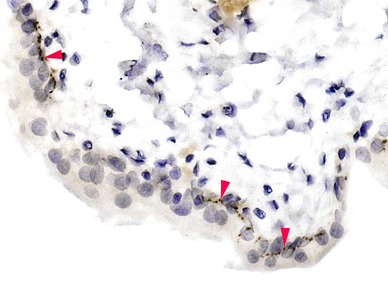
Red arrowheads indicate positive nerve fibres within the urothelium.
An example of a situation where IF would give far better visualisation of the intra-urothelial nerve fibres.
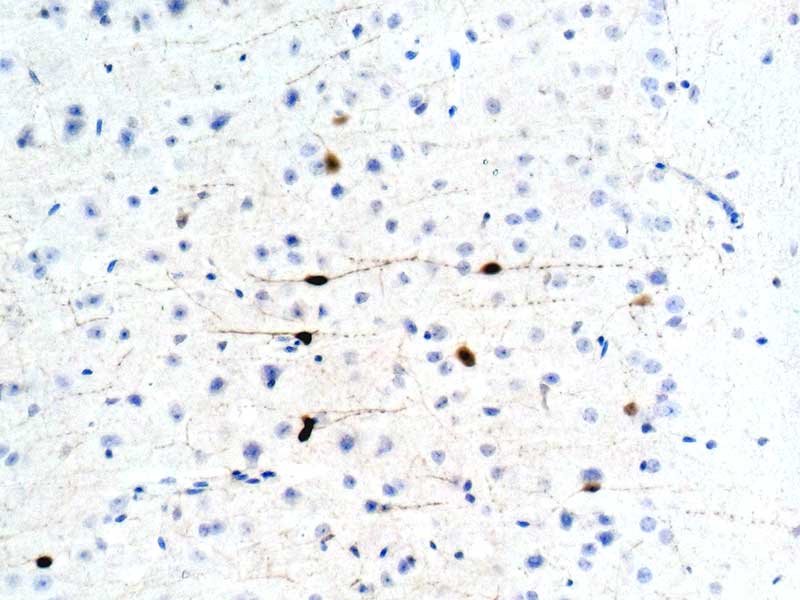
I do not know if this is specific
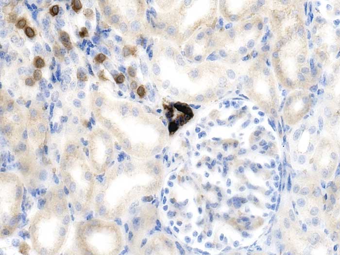
Occasional cells in the collecting tubules are positive.
The DCT that intimately associates with the vascular pole( part of the JGA) is strongly positive ( not just the Macula densa).
Comments please?
I cannot find confirmation that this result is specific.
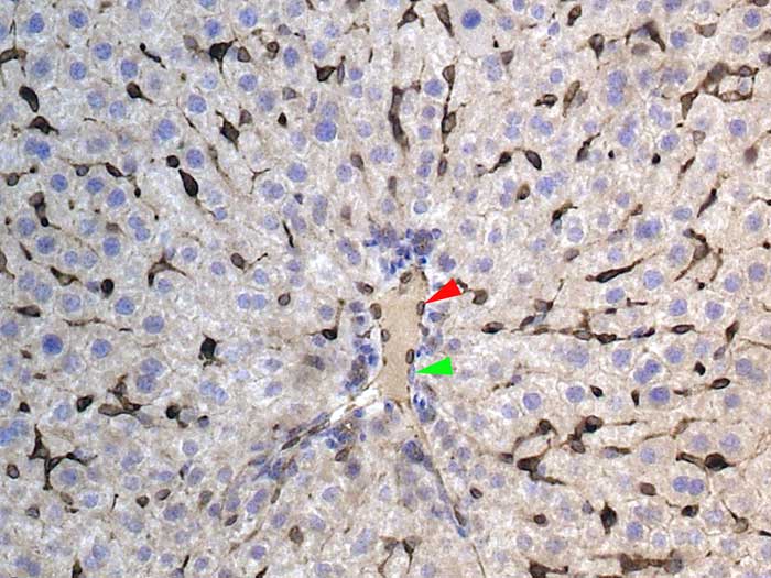
Kupffer cells are positive
Cells lining the central vein are also positive ( red a’head).
Green a’head indicates a negative lining cell.
Are they both endothelial cells?
I would be grateful for your considered opinion.
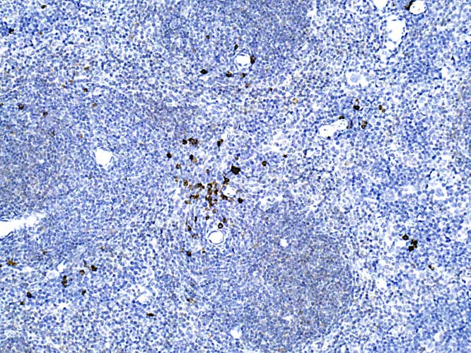
Many strongly positive neutrophils are seen ( in the artery, most of which are adherent to the endothelium).
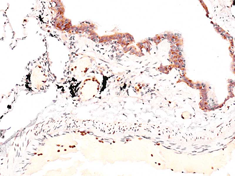
Bronchiolar epithelial lining cells also have cytoplasmic positivity.
NB: The black pigment is most likely to be carbon.
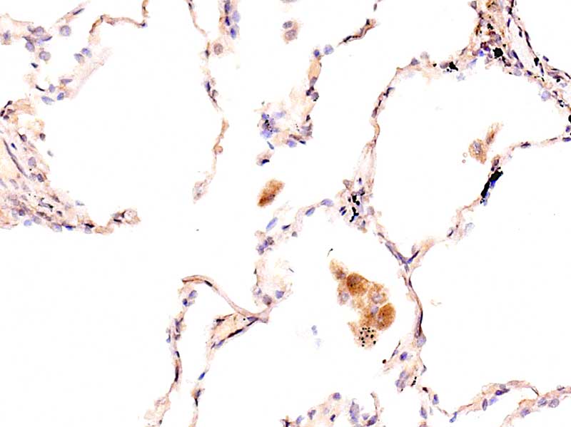
In this image positive macrophages are seen.
Unlike when immunodetecting Iba1, not all macrophages are positive for Cathelicidin.