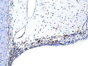
Progenitor olfactory bulb interneurons and endothelial cells are positive.
Lateral ventricle.
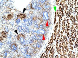
Coloured arrowheads indicate positivity:
red for a linear positivity around many tubules ( ? basal lamina?); black for developing Glomeruli; green for myotubes T/S.
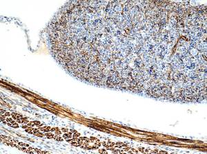
Interstitial cells in developing spleen ( upper right) are positive.
Lower left shows mostly primary myotubules in T/S and L/S that are positive.
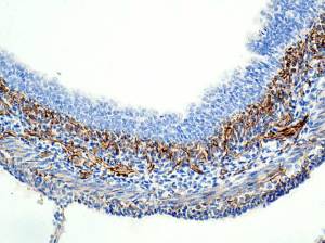
Developing bladder. Stromal cells of the lamina propria are positive.
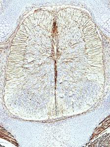
T/S developing spinal cord. The radial patterning of Nestin-positive fibres appears to be similar to that of GFAP in adult spinal cord.
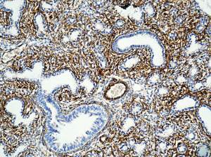
Developing lung shows positivity in stromal cells and also ? basal laminae?
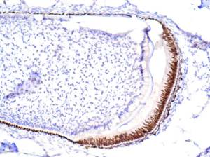
Hmmm…are ameloblast nuclei positive and odontoblast nuclei negative?
Comments welcome!
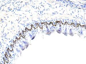
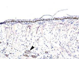
Buccal epithelium. Filiform papillae only?
Positivity confined to basal epithelial nuclei?
 .
.
Lingual surface
Arrowhead indicates nerve fibre positivity.
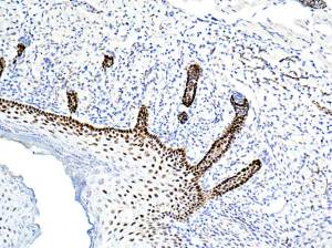
Most layers of epidermal nuclei are positive.
Hair follicle nuclei the same .
Upload Images for Histonet