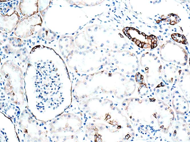
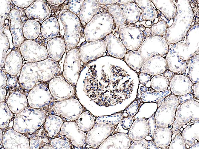
Cortex: most pronounced positivity is seen in collecting tubule/duct epithelia.
Occcasional PCT/DCT epithelial cells are positive. Parietal cells of Bowman’s capsule are also positive.
Cortex: most pronounced positivity is seen in collecting tubule/duct epithelia.
Occcasional PCT/DCT epithelial cells are positive. Parietal cells of Bowman’s capsule are also positive.
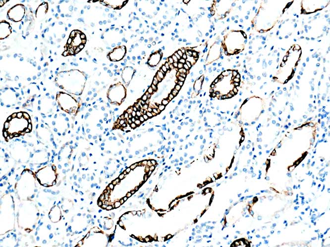
Positivity using this ab ( anti K5/8) appears to be confined to collecting duct and Bowman’s capsule parietal epithelium.

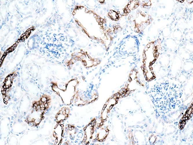
Renal papilla showing positive simple cuboidal ( collecting ducts) and transitional epithelia ( urothelium covering of the papilla).
This reagent is stated as being anti Keratins 5/8.
However, as K5 is found mainly in basal epithelial cells of epidermis ( I think) this collecting duct positivity must be for K8.
Apart from transitional cells, no other epithelia in kidney are positive.

Top: CD68
Middle: Iba1
Bottom: MMP9
Rat cannulation injury site.
Comments welcome re the fact that MMP9 appears to be positive in a subset of CD68 positive macrophages.
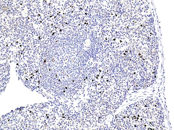
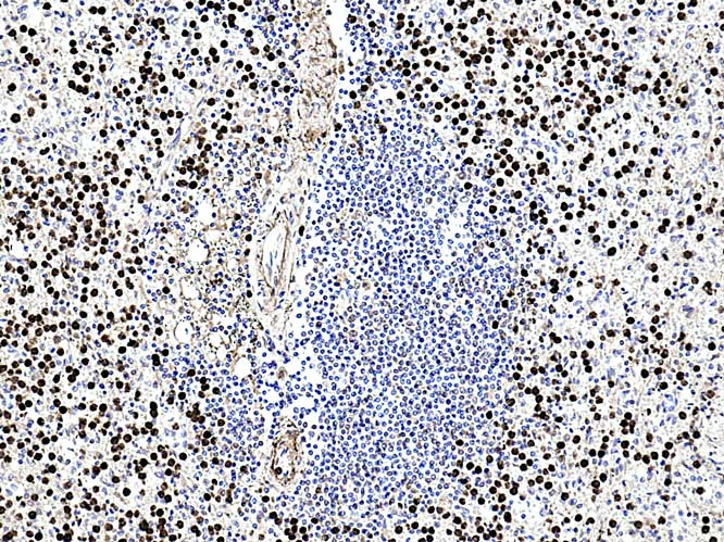
Antibody should be further diluted ( 1/3K used). Positivity appears confined to macrophages of the red pulp sinusoids.
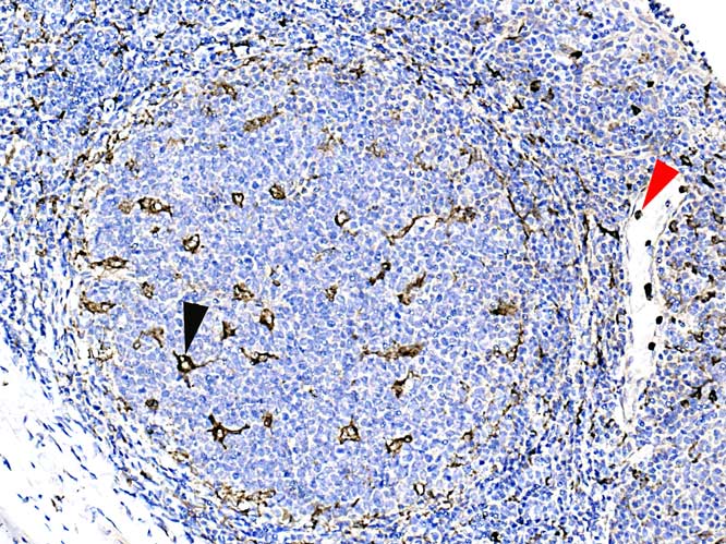
Germinal centres show positive dendritic macrophages ( black arrowhead)
Rounded, smaller +ve cells are seen in the high endothelial venules ( red arrowhead) Are these neutrophils?
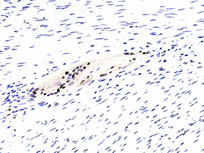
In the muscularis externa positivity is seen only in the nuclei of
?satellite cells? associated with the ganglion cell bodies of
Auerbach’ s plexus