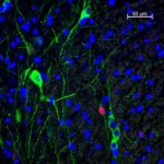
In this field, two dopaminergic cells ( SNc) have FOS +ve nuclei
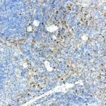
Should be further diluted ( I used 1/2K)
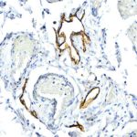
Positivity is seen only in the endothelial cells lining the lymphatic vessels.
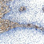
Positivity is confined to the collagen fibres of the interstitial tissues. No Biglycan is detected in the mucosal epithelium
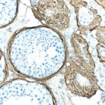
Positivity is seen within the collagen fibres of the interstitial tissue
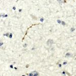
Image of frontal cortex shows occasional small nerve fibres showing a beaded positivty

Predominantly expressed in the hypothalamus.
However, there is also a noticable positivity in the brainstem
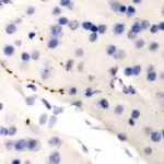
Small, scattered and seemingly isolated fibres give a beaded positivity ( cortex)
Upload Images for Histonet