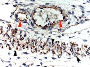
T/S of developing skeletal muscle shows positivity at junction of primary/secondary myotubes ( black arrowheads).
Red arrowheads indicate blood vessel smooth muscle positivity.
I have no idea if this is “true” positivity. I welcome comments.
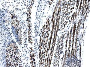
At this stage one can see positive primary myotubes in L/S and T/S.
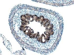
Strangely/interestingly, epithelium is strongly positive.
Specific?
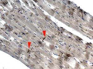
Not the best quality section.
I have stopped down the substage lens to induce refraction so that striations can be seen.
Red arrowheads indicate Z-discs, which are intensely positive ( antibody should be further diluted).
I do not know if this positivity is specific; I welcome comments.
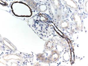
In mouse, this antibody appears to be specific for smooth muscle.
There is however, a moderate positivity in the microvilli of the PCT.
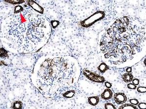
Cortex: obvious positivity of podocytes and distal convoluted tubule at site of Macula densa ( red arrowhead). Not sure why many tubules are negative.
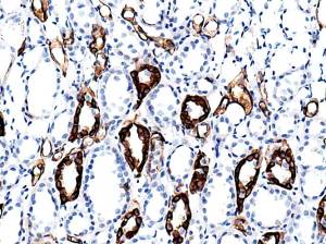
Medulla. Seems to me that Loop of Henle epithelium is positive. Collecting ducts are negative.
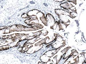
Bronchial epithelium is positive.
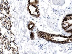
Strong positivity in bronchiolar and alveolar epithelium.
Upload Images for Histonet