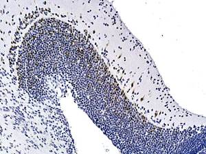
Not sure if this is good/representative positivity.
I would expect more neurones to be positive.
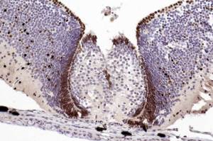
Skin at base of specimen shows melanin pigment, not DAB.
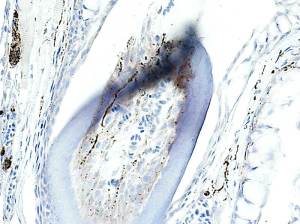
Nerve fibres within the tooth pulp are clearly seen. Sure, the dentine is disrupted ( due to the aggressive nature of HIER).
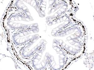
Submucosal/myenteric plexi and individual fibres in lamina propria are very well demonstrated.
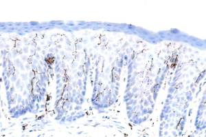
Nerve fibres in pharyngeal epithelium.
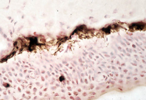
Intense melanocyte positivity that extends, partially , into the processes that insert melanin into keratinocytes.
Upload Images for Histonet