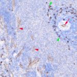
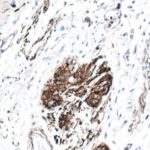
The datasheet states that it has been reported to x-react with eNOS and also nNOS. On FFPWS after Citric acid HIER I concur. Submitted images shows clear endothelial positivity, I am not sure what the heavily positive cells in the marginal zone of the white pulp are ( also some around high endothelial venules).
All posts by Carl Hobbs
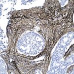
COLLAGEN TYPE III IN HUMAN OVARIAN CARCINOMA: FFPWS USING ABCAM AB7778
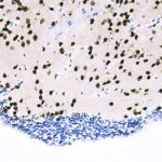
NEUN IN MOUSE BRAIN : Free-floating fixed section USING ABCAM AB177487
NEUN IN MOUSE BRAIN : FFPWS USING ABCAM AB177487
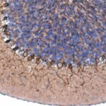
ATG7 IN mouse cerebellum : FFPWS USING ABCAM AB133528
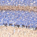
ATG7 IN rat cerebellum : FFPWS USING ABCAM AB133528
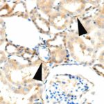
ATG7 IN marmoset kidney : FFPWS USING ABCAM AB133528
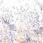
ATG7 IN HUMAN kidney : FFPWS USING ABCAM AB133528
ATG7 in human colon: FFPWS using abcam ab133528
I do not know the specificity of these results. Abcam datasheet includes “…..plays also a key role in the
maintenance of axonal homeostasis, the prevention of axonal degeneration…”
Abcam datasheet does not state IHC-P reactivity.
Positivity is confined to neuronal cytoplasm/processes in central and peripheral nervous system.
Image shows such positivity in nerve fibres and also Auerbach’s plexus neurone cell cytoplasm
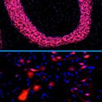
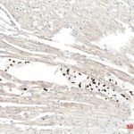
Human nuclei in mouse heart : FFPWS USING Abcam ab190710
I did the initial histology and IHC for a student to establish the “best marker” to identify the cells if they were found in the heart. Once we agreed that ab190710 gave the best results ( I also tested anti TFAM, MTCO2 and STEM121 : all human -specific) the student carried on without aid. I also probed with anti GFP as the progenitor cells were labelled with GFP. They all worked but gave an inferior signal:noise
The PhD student: I injected human cardiac progenitor cells (c-kit+, CD31-) isolated from human heart tissue into NSG mice.
