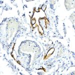
Positivity is seen only in the endothelial cells lining the lymphatic vessels.
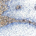
Positivity is confined to the collagen fibres of the interstitial tissues. No Biglycan is detected in the mucosal epithelium
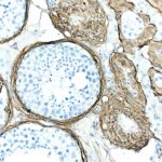
Positivity is seen within the collagen fibres of the interstitial tissue
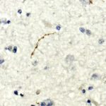
Image of frontal cortex shows occasional small nerve fibres showing a beaded positivty

Predominantly expressed in the hypothalamus.
However, there is also a noticable positivity in the brainstem
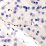
Small, scattered and seemingly isolated fibres give a beaded positivity ( cortex)
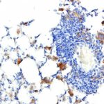
Inflamed lung showing positive macrophages. Some of them have fused to form giant cells
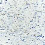
Above the hippocampal CA1 region: the corpus callosum runs from LtoR along the bottom. Concentrated positivity is seen in hypothalamus but, as seen here there is scattered small fibre punctate-positivity in many other areas.
Upload Images for Histonet