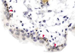
Red arrowheads indicate positive nerve fibres within the urothelium.
An example of a situation where IF would give far better visualisation of the intra-urothelial nerve fibres.
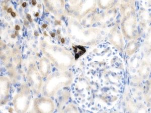
I do not know if this is specific
Occasional cells in the collecting tubules are positive.
The DCT that intimately associates with the vascular pole( part of the JGA) is strongly positive ( not just the Macula densa).
Comments please?
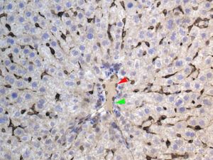
I cannot find confirmation that this result is specific.
Kupffer cells are positive
Cells lining the central vein are also positive ( red a’head).
Green a’head indicates a negative lining cell.
Are they both endothelial cells?
I would be grateful for your considered opinion.
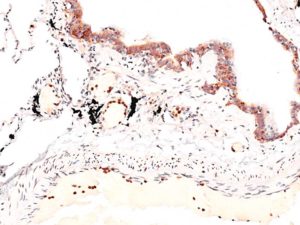
Many strongly positive neutrophils are seen ( in the artery, most of which are adherent to the endothelium).
Bronchiolar epithelial lining cells also have cytoplasmic positivity.
NB: The black pigment is most likely to be carbon.
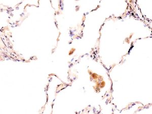
In this image positive macrophages are seen.
Unlike when immunodetecting Iba1, not all macrophages are positive for Cathelicidin.
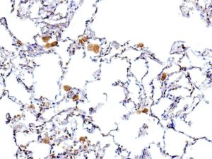
Positive macrophages.
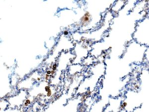
Positive macrophages
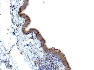
Positive bronchiolar epithelium
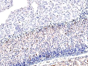
Adrenal gland from a different mouse; no medullary cell positivity.
Upload Images for Histonet