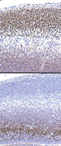
T/S through frontal cortex ( mid-sagittal)
Top image TBR1.
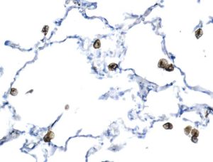
Lung macrophages are positive.
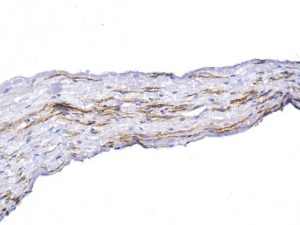
Not shown but, to the right is the spinal cord ( dorsal).
Some of the nerve fibres of the root are positive for CGRP.
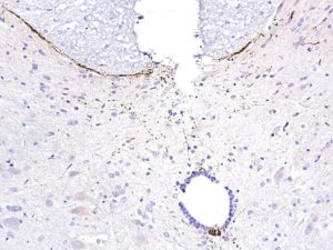
Mid-cord: a single positive fibre traverses the cord. Also, there is a ?positivity within some cells of the canal.
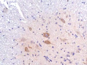
Ventral horn: punctate cytoplasmic positivity in motor neurones.
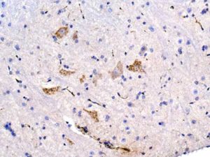
Ventral horn showing punctate cytoplasmic positivity in motor neurones.
Sure, there is also positivity in capillaries ( IgG).
This specimen was immersion-fixed.
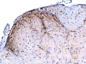
Should be further diluted ( I used 1/2K).
Gives the same expression pattern as other Abs that also work in Pwax sections, under same conditions.
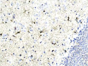
Strong positivity in microglia.
Upload Images for Histonet