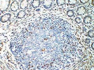
Peyers patch in submucosa
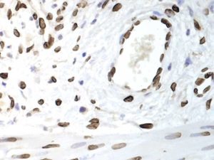
Arteriole in submucosa,
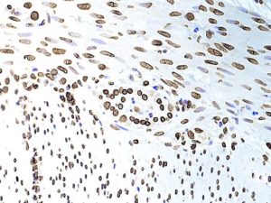
Muscularis externa of jejunum ) Auerbach’s plexus seen, centrally.
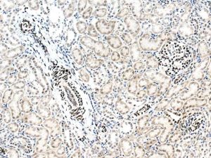
Antibody should be further diluted to remove the cytoplasmic stasining.
Most intense Lamin positivity is seen in smooth muscle nuclei of the blood vessels and the podocytes of the glomerulus.
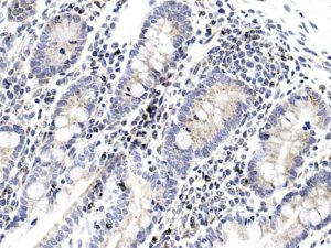
Mucosa area. Should I have expected to see strong positivity in enterocytes?
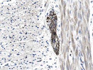
Cells/fibres of Aurbach’s plexuses are particularly enriched ( if specific, of course)
Upload Images for Histonet