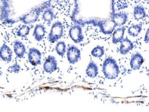
Very thin muscularis externa but, cells and fibres of enteric nerve plexuses are clearly positive.
Neuroendocrine cells ( I assume) are better shown here compared to the colon.
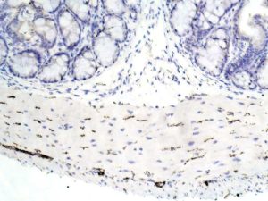
Positivity is seen in fibres and cell bodies of enteric plexuses.
I note individual cell positivity amongst the enterocytes….neuroendocrine cells?
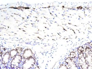
A slight enterocyte cytoplasmic positivity that should disappear on further dilution of primary Ab ( a little too strong).
Note high levels of protein in fibres and cell bodies of both enteric plexuses.
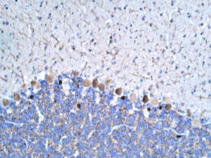
Cerebellum
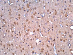
Frontal cortex
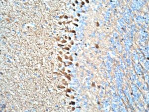
Olfactory bulb
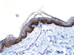
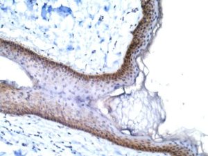
Average: appropriate region is positive but not as striking as Covance ab on mouse tissue.
( note that Covance ab is negative for human, however).
Upload Images for Histonet