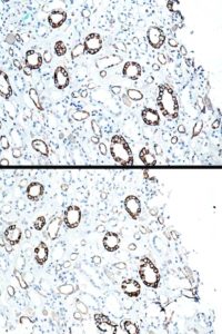
BD upper image. There appears to be an identical pattern of positivity.
Medulla of human kidney: FFPWS.
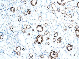
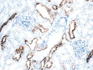
Medulla: strong immunostaining of collecting duct and Loop of Henle epithelium.
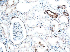
Cortex: most pronounced positivity is seen in collecting tubule/duct epithelia.
Occcasional PCT/DCT epithelial cells are positive. Parietal cells of Bowman’s capsule are also positive.
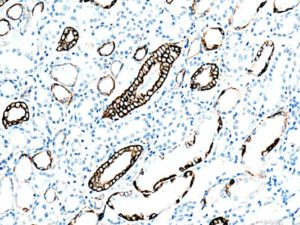
Positivity using this ab ( anti K5/8) appears to be confined to collecting duct and Bowman’s capsule parietal epithelium.
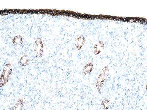
Renal papilla showing positive simple cuboidal ( collecting ducts) and transitional epithelia ( urothelium covering of the papilla).
This reagent is stated as being anti Keratins 5/8.
However, as K5 is found mainly in basal epithelial cells of epidermis ( I think) this collecting duct positivity must be for K8.
Apart from transitional cells, no other epithelia in kidney are positive.
Upload Images for Histonet