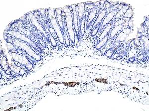
Auerbach’s plexus is easily seen.
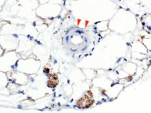
Nerve bundles and individual nerve fibres ( red) appear positive.
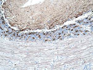
A poorly fixed specimen.
Is the positivity “real”?
It differs from mouse in that Purkinje cells are not positive.
However, Basket cells are intensely positive.
As are the peripheries of Ganglion cell glomeruli ( same as mouse).
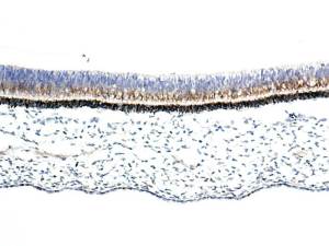
Seems to me that the positivity is confined to the cell bodies of the rods and cones?
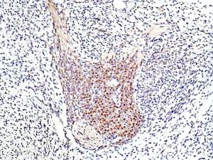
Very different positivity: seemingly Golgi in ganglion cells ?
Also, there is a moderate fibre positivity.
Upload Images for Histonet