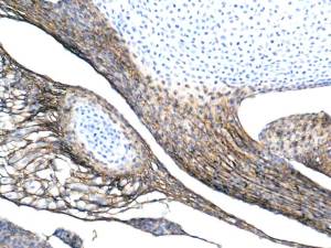
Collagen Type I in developing tendons ( the bone is still cartilage).
Is this positivity specific?
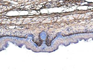
Collagen Type I is positive.
Intense positivity is seen in the sub -basal lamina region.
Comments are wanted.
Sure…the Ab solution should be further diluted.
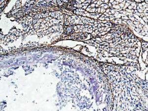
Is this specific?
I see only collagen Type I fibre positivity.
Lower left is a developing rib so osteoblasts and osteoclasts are present; none are positive.
I welcome opinions.
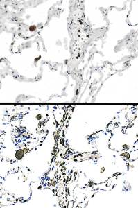
MMP9: upper image. Lower image is of Iba1. Indicates that MMP9 is in a sub-population of macrophages?
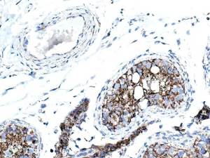
Seminiferous tubule and Leydig cells are enriched.
Strong, fine punctate positivity is also seen in stromal fibroblasts and smooth muscle cells of the arteriole.
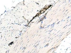
Auerbach’s ganglion cells show a very strong membraneous positivity.
Upload Images for Histonet