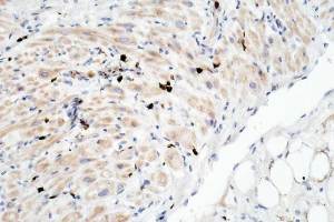
A slight positivity in myofibre cytoplasm; this may/not be due to insufficient dilution of primary ab.
What appears to be an occasional intense nuclear positivity is seen.
This appears to be in cells between myofibres
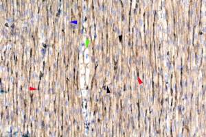
Coloured arrowheads indicate positivity.
Intense blood vessel positivity( red).
Moderate cardiomyocyte membrane positivity( blue)
Moderate intercalated disc positivity ( black)
A moderate cardiomyocyte positivity…specific??
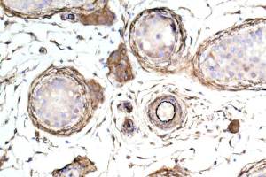
hmmm…not at all sure about this pattern of positivity.
However, human heart gives a positivity that seems to be specific.
Intense arteriole endothelial cell positivity. The connective tissue surrounding each seminiferous tubule is also positive.
Cytoplasm of Fibrocytes and Leydig cells are also positive.
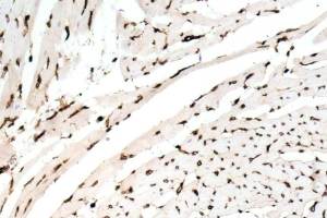
Intense endothelial cell positivity. Cardiomyocyte membrane positivity is present though is pale, compared to other species.
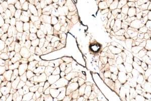
Intense endothelial cell positivity.
A distinct cardiomyocyte membrane positivity is seen.
At high power ( not clear in this image) the cytoplasmic positivity of the myocytes appear to delineate myofibrils.
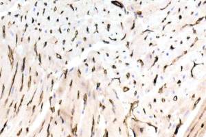
Intense endothelial cell immunostaining. A slight cardiomyocyte cytoplasm positivity is, imho, artefact.
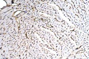
Intense endothelial positivity. Myocyte membranes are positive in some specie but, not others ( see human image).
Upload Images for Histonet