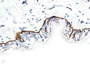
Does not work using HIER. Use Proteinase K digestion.
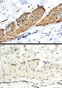
Upper image shows delineation of smooth muscle cells’ basal laminae. Lower image shows no staining after probing with anti Collagen VII.
Proteinase K digestion required. Both Abs negative on FFPWS +/- HIER.
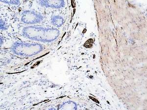
My thanks to the very kind Dr Bozkurt for all my ” exotic ” tissues.
A slight ( non-specific?) positivity in the muscularis externa ( also positive when using Ultraclone rb polyclonal).
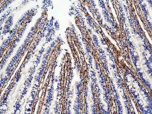
Interesting positivity in the lamina propria of the villi; are they endothelial cells?
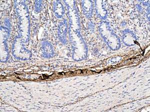
The dark brown positivity seen in the submucosa is artefactual : the collagen is disrupted.
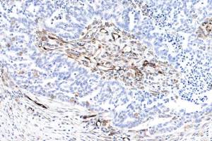
Tumour cells are negative. Endothelial and stromal cells are positive.
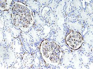
Pattern of positivity identical to that seen in three different antibodies tested by the HPA.
Parietal cells of the Bowman’s capsule; some cells within the glomerulus and cells within the interstitium or they are endothelial cells.
Upload Images for Histonet