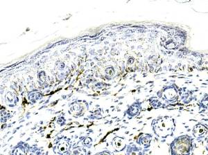
Fine fibres are seen but, as expected, they are easier to visualise when using IF.
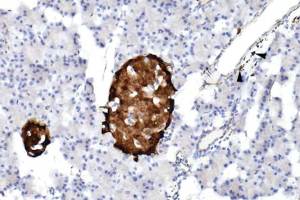
Most of the IofL cells are positive. A few are remarkably negative.
Black arrowheads indicate positivity seen in fine nerve fibres running along a blood vessel.
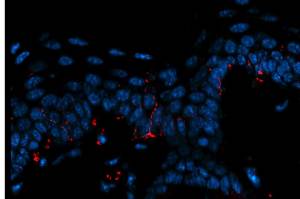
Detection of intra-epidermal nerve fibres: anti Rb Alexa 594. 8 micron section.
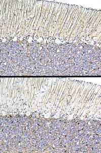
Upper image using Sigma G3893 and lower image using Abcam ab4674
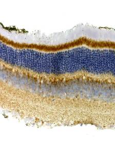
Or…is this ?
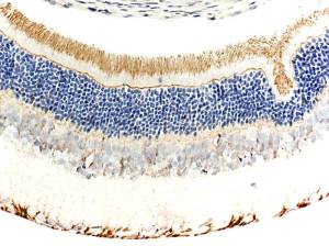
Is this a “correct” pattern of positivity?
Upload Images for Histonet