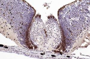
Skin at base of specimen shows melanin pigment, not DAB.
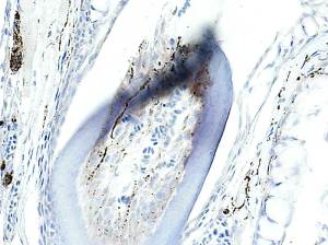
Nerve fibres within the tooth pulp are clearly seen. Sure, the dentine is disrupted ( due to the aggressive nature of HIER).
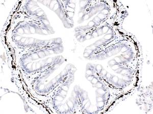
Submucosal/myenteric plexi and individual fibres in lamina propria are very well demonstrated.
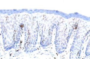
Nerve fibres in pharyngeal epithelium.
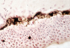
Intense melanocyte positivity that extends, partially , into the processes that insert melanin into keratinocytes.
Upload Images for Histonet