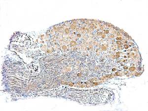
Similar distribution/intensity pattern to that seen in mouse DRG, under identical conditions.
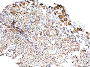
Strong cytoplasmic positivity in ganglion cells ( variable intensity) and nerve fibres.
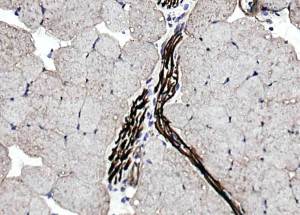
Myelinated nerve tracts showing intense positivity compared with skeletal muscle fibres.
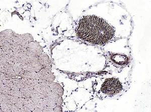
A lighter positivity in skeletal muscle but, a very intense positivity in myelinated nerve fibres and also arteriolar smooth muscle.
Smooth muscle of venules appears to be negative.
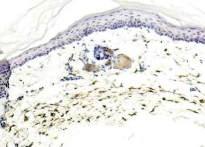
Ignore the sebaceous gland positivity.
Upload Images for Histonet