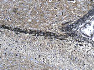
NCAD is enriched in svz and migrating olfactory interneurons ( in the RMS).
Note that capillary positivity is due to plasma IgGs and not NCAD.
( the brain was not perfused)
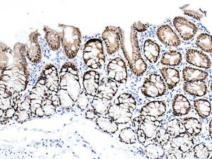
There is a moderate cytoplasmic positivity.
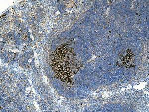
Immunostaining intensity is far too strong as I used a dilution factor that is optimal for my homemade DAB solution but, I developed using a commercial, enhanced- DAB kit.
However, an interesting pattern of positivity.
I would appreciate comments/explanations.
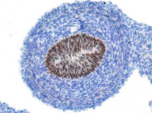
I am not yet familiar with the distribution pattern of this protein. However, published data states that this protein is present in the nuclei of gut mucosal epithelium ( embryonic and adult).
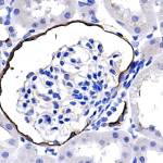
Parietal cells of the glomerulus and also distal cells of the Macula densa ( DCT) are strongly positive.
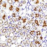
Medulla: given that the PCTs’ surface epithelium shows strong positivity ( in the cortex) I assume that the irregular intense positivity here is the descending ( thick) part of the Loop of Henle. Most other tubular structures ( whether collecting ducts/thin loops of Henle) show a moderate surface epithelial positivity.
Upload Images for Histonet