Abcam state that this ab is only human-reactive.
Sure, I am not familiar with this protein.
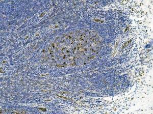
Cytoplasmic positivity is confined to three areas: what appear to be mononuclear phagocyte cells within an activated nodule; high endothelial venules ( could be migrating macrophages); large cells within internodular areas.
Comments would be appreciated.
Not an “impressive” positivity but, immunostaining carried out at same time as all other submitted images.
Does Atrogin 1 have a developmental function?
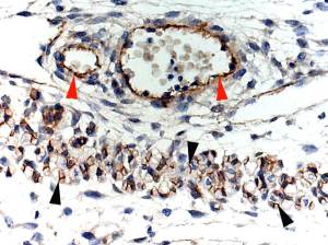
T/S of developing skeletal muscle shows positivity at junction of primary/secondary myotubes ( black arrowheads).
Red arrowheads indicate blood vessel smooth muscle positivity.
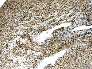
I have no idea if this is “true” positivity. I welcome comments.
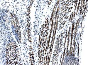
At this stage one can see positive primary myotubes in L/S and T/S.
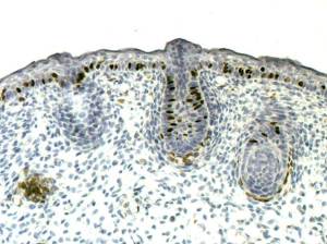
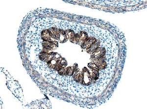
Strangely/interestingly, epithelium is strongly positive.
Specific?
Not the best quality section.
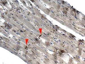
I have stopped down the substage lens to induce refraction so that striations can be seen.
Red arrowheads indicate Z-discs, which are intensely positive ( antibody should be further diluted).
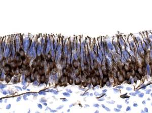
I do not know if this positivity is specific; I welcome comments.
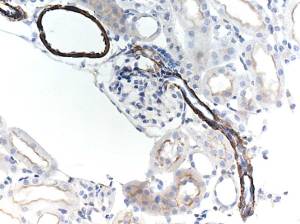
In mouse, this antibody appears to be specific for smooth muscle.
There is however, a moderate positivity in the microvilli of the PCT.
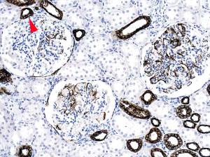
Cortex: obvious positivity of podocytes and distal convoluted tubule at site of Macula densa ( red arrowhead). Not sure why many tubules are negative.
Posts navigation
Upload Images for Histonet
