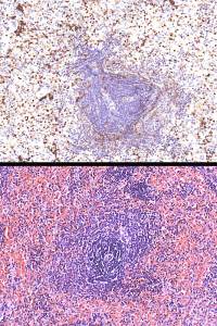
Upper image shows positivity in clusters of cells , mostly within the red pulp ( there are some positive cells within the white pulp, however).
Lower image is a H&E to demonstrate that mature rbcs are negative for this antibody ( the pink cells with no nuclei).