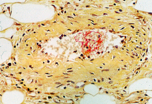
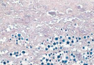
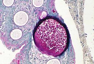
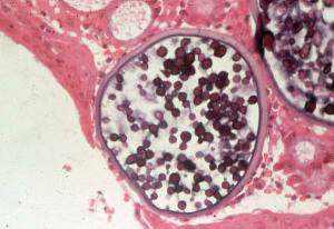
An early clot, noticed by chance.
Images
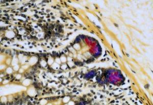
Paneth cell granules in human jejunum: FFPWS using Lendrum’s Phloxine Tartrazine.
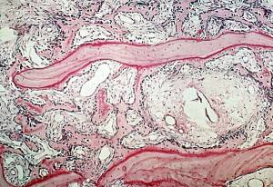
Osteioid seams in developing bone of human tumour: FFPWS using PAS technique ( after decalcification).
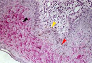
Glycogen in human skin: FFPWS using PAS technique
Predominantly in stratum spinosal cells. Note that glycogen is assymetrically distributed within each cell ( black arrowhead). This is because of the unidirectional penetration/flow of fixative when a specimen is placed in fixing fluid without agitation: the glycogen is pushed ahead of the wave of fixing fluid until it cannot go further because of the cell membrane.
Melanin within melanocytes is indicated by a yellow arrowhead. After transportation into cells of the basal layer ( red arrowhead), melanin forms a cap over the nuclei ( protecting DNA from UV light…up to a point!).
