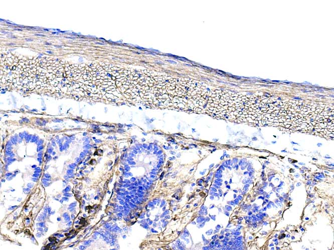
Positivity appears restricted to basal laminae ( particularly evident in the T/S presentation of smooth muscle cells of the inner layer of the muscularis externa : this is a longitudinal orientation of the specimen so, the inner circular layer shows as T/S)
The intense positivity seen in cells within the lamina propria is due to endogenous IgG ( Plasma cells) as I forgot to use rat-adsorbed anti mouse IgG secondary antibody.