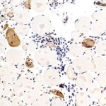
Biopsy from a patient suffering from Polymyositis. Accumulations of lymphocytes can be seen (intensely-stained rounded, blue nuclei)
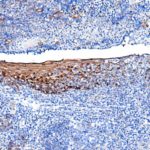
I do not know why ADH would be present in mucosal tissue but HPA show identical pattern with their enhanced orthogonal-validated abs.
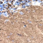
Cerebellum: strong positivity is seen in molecular layer and Glomeruli of Granule cells. However, most intense positivity in seen in microglia.
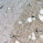
Dentate nucleus. Most intense positivity is seen within microglia.
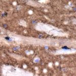
Striatum: Pia is heavily stained ( unlike nerve fibre tracts) but microglia are the most intensely +ve.
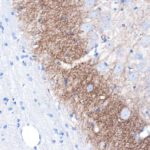
Hippocampus CA2 region ( to the left is corpus callosum-negative)
Upload Images for Histonet