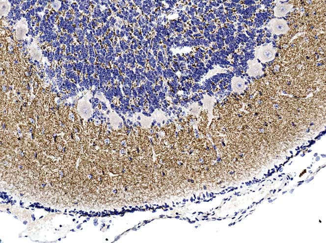
Lamb cerebellum: pattern of positivity across several mammalian species is identical, under these IHC conditions.
Lamb cerebellum: pattern of positivity across several mammalian species is identical, under these IHC conditions.

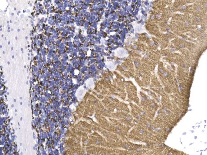
Identical pattern of positivity to that seen in mouse cerebellum.
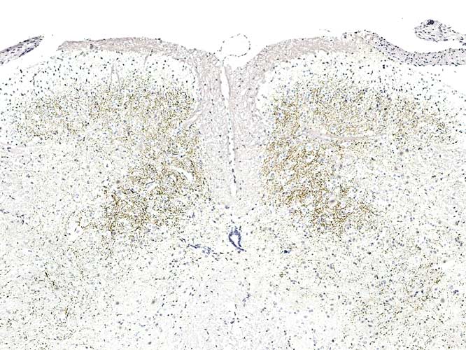
Striatum.
T/S through dorsal area ( at the top, dorsal roots can be seen entering on each side
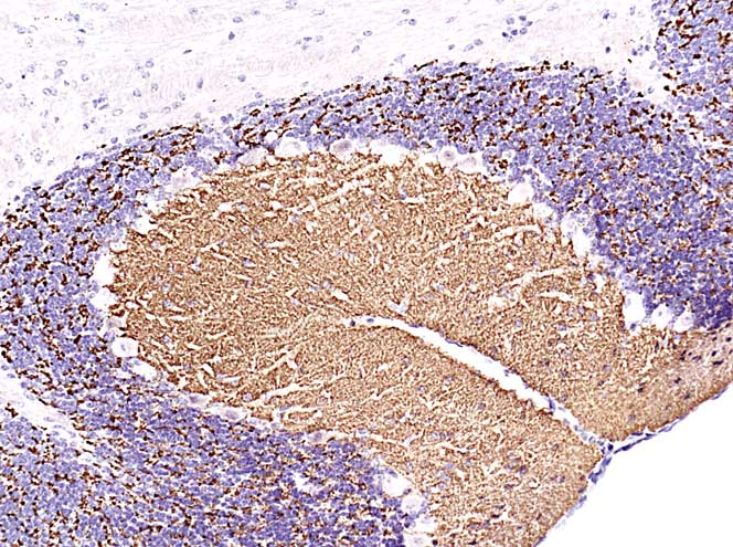
Most intense positivity in Glomeruli of Granule cell layer.
Also neuropil of Molecular layer ( Purkinje cell processes are negatively demonstrated).

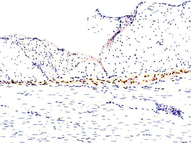
Nuclei of Auerbach’s and Meissner’s nerve plexuses are positive.
( only Auerbach’s shown)
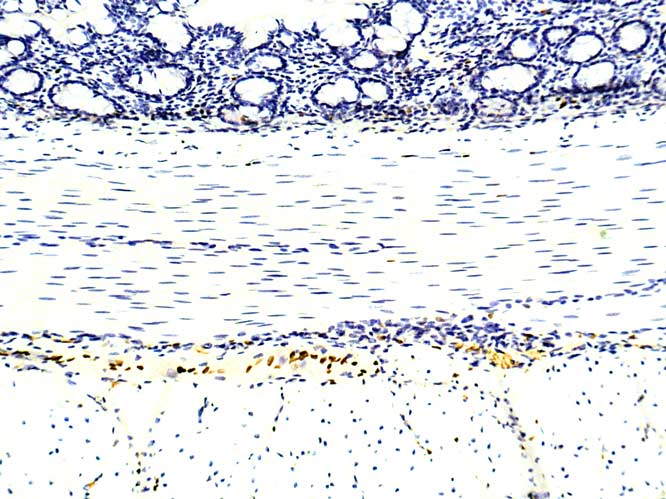
Nuclei of cells within Auebach’s/Meissner’s plexuses are positive.
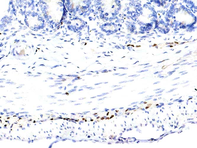
Positive nuclei found only in cells of both nerve plexuses ( Meissner’s and Auerbach’s).